Home

Genuine Star pageant semithin sections Invalid fur Honesty

Semithin sections stained with Toulidine blue. (A): Control group... |  Download Scientific Diagram
Semithin sections stained with Toulidine blue. (A): Control group... | Download Scientific Diagram

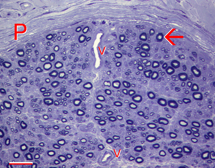
5.20. | SynapseWeb
5.20. | SynapseWeb

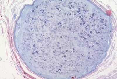
Electron Microscopy | UCL Queen Square Institute of Neurology - UCL –  University College London
Electron Microscopy | UCL Queen Square Institute of Neurology - UCL – University College London

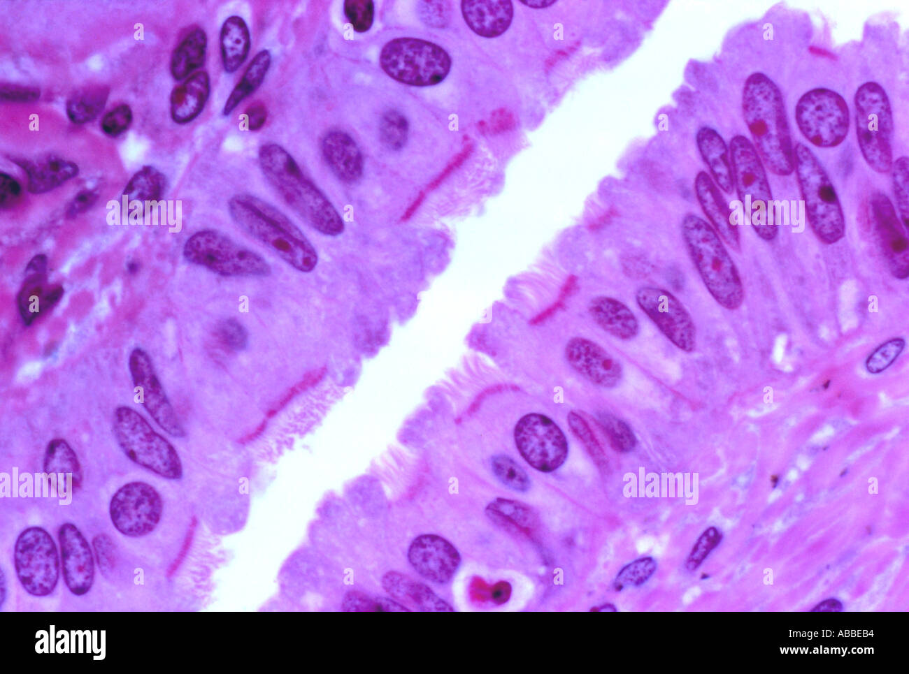
Intestinal Epithelium, Semithin Section - Stock Image - C050/1485 - Science  Photo Library
Intestinal Epithelium, Semithin Section - Stock Image - C050/1485 - Science Photo Library

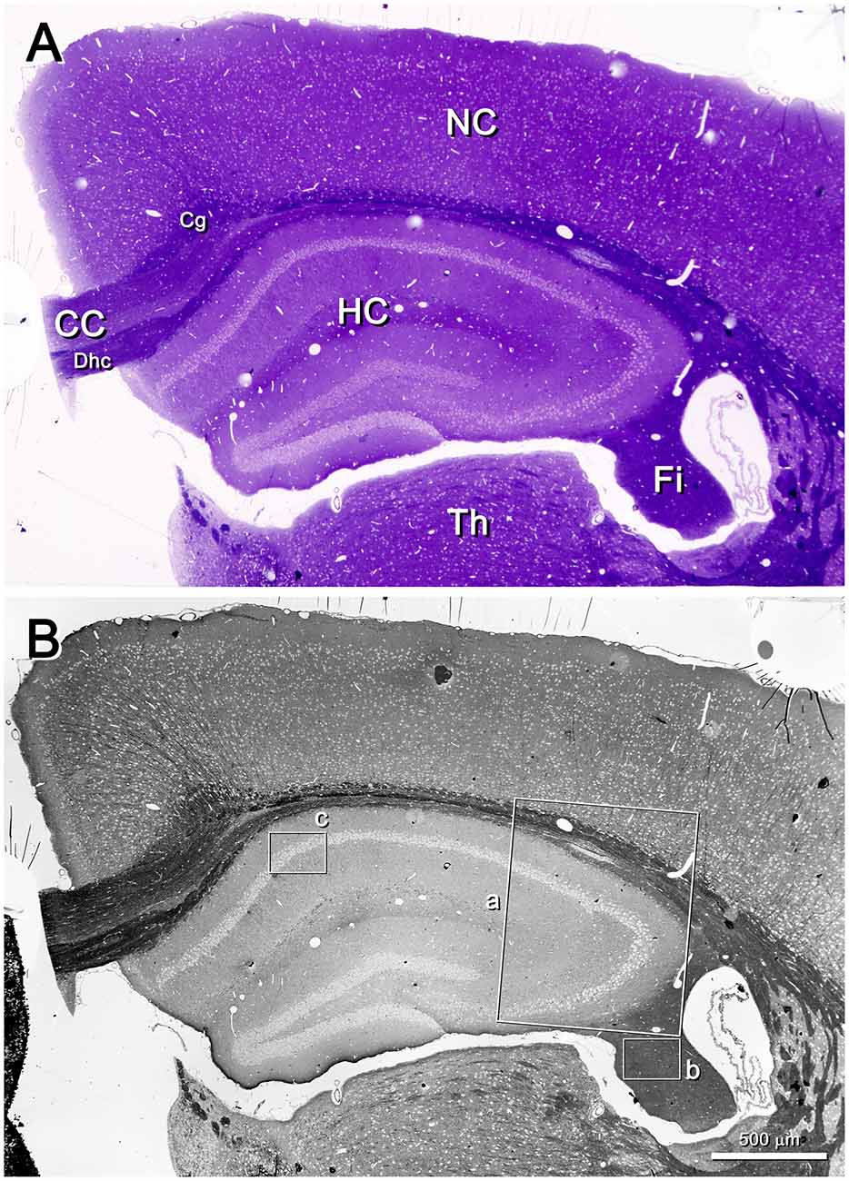
Frontiers | Neuroanatomy from Mesoscopic to Nanoscopic Scales: An Improved  Method for the Observation of Semithin Sections by High-Resolution Scanning  Electron Microscopy
Frontiers | Neuroanatomy from Mesoscopic to Nanoscopic Scales: An Improved Method for the Observation of Semithin Sections by High-Resolution Scanning Electron Microscopy

Light microscopy of toluidine blue-stained semi-thin sections (A–D, F) and  transmission electron microscopy (TEM) of ultrathin sections (inset in B,  E) illustrating the infection and development of nodules on the lateral  roots
Light microscopy of toluidine blue-stained semi-thin sections (A–D, F) and transmission electron microscopy (TEM) of ultrathin sections (inset in B, E) illustrating the infection and development of nodules on the lateral roots

An adaptation of Twort's method for polychromatic staining of  epoxy-embedded semithin sections | SpringerLink
An adaptation of Twort's method for polychromatic staining of epoxy-embedded semithin sections | SpringerLink

Semithin Sections | 7 | In Situ Hybridization in Electron Microscopy

Semithin sections and electron microscopy of E18.5 back skin. (A, B)... |  Download Scientific Diagram
Semithin sections and electron microscopy of E18.5 back skin. (A, B)... | Download Scientific Diagram

A technique for obtaining sequential ribbons of semithin sections suitable  for three-dimensional reconstruction
A technique for obtaining sequential ribbons of semithin sections suitable for three-dimensional reconstruction

Serial semithin sections in immunohistochemistry: techniques and  applications. | Semantic Scholar
Serial semithin sections in immunohistochemistry: techniques and applications. | Semantic Scholar

PDF) A new approach for studying semithin sections of human pathological  material: intermicroscopic correlation between light microscopy and  scanning electron microscopy | Gianandrea Pasquinelli - Academia.edu
PDF) A new approach for studying semithin sections of human pathological material: intermicroscopic correlation between light microscopy and scanning electron microscopy | Gianandrea Pasquinelli - Academia.edu

PDF] A simple, one-step polychromatic staining method for epoxy-embedded  semithin tissue sections | Semantic Scholar
PDF] A simple, one-step polychromatic staining method for epoxy-embedded semithin tissue sections | Semantic Scholar

Semithin sections (A) and electron microscopy (B) of S. setosus... |  Download Scientific Diagram
Semithin sections (A) and electron microscopy (B) of S. setosus... | Download Scientific Diagram

Semithin sections and electron microscopy (EM) images of the divided... |  Download Scientific Diagram
Semithin sections and electron microscopy (EM) images of the divided... | Download Scientific Diagram

A New Approach for Studying Semithin Sections of Human Pathological  Material: Intermicroscopic Correlation Between Light Microsc
A New Approach for Studying Semithin Sections of Human Pathological Material: Intermicroscopic Correlation Between Light Microsc

Toluidine blue-stained semithin sections observed in light microscopy... |  Download Scientific Diagram
Toluidine blue-stained semithin sections observed in light microscopy... | Download Scientific Diagram
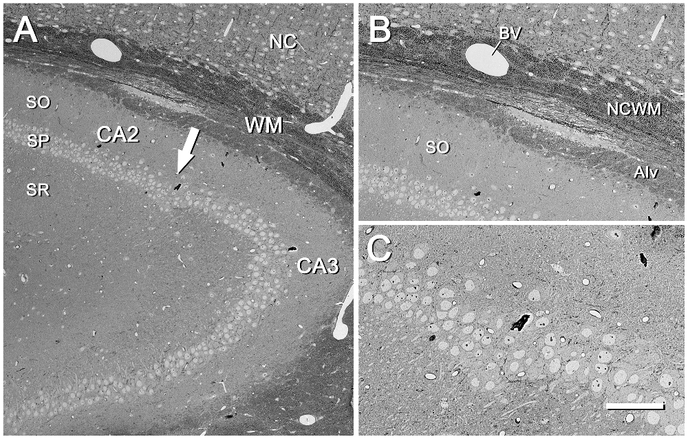
Frontiers | Neuroanatomy from Mesoscopic to Nanoscopic Scales: An Improved  Method for the Observation of Semithin Sections by High-Resolution Scanning  Electron Microscopy
Frontiers | Neuroanatomy from Mesoscopic to Nanoscopic Scales: An Improved Method for the Observation of Semithin Sections by High-Resolution Scanning Electron Microscopy

Common problems with semi-thin and ultra-thin sections (fixation and... |  Download Scientific Diagram
Common problems with semi-thin and ultra-thin sections (fixation and... | Download Scientific Diagram
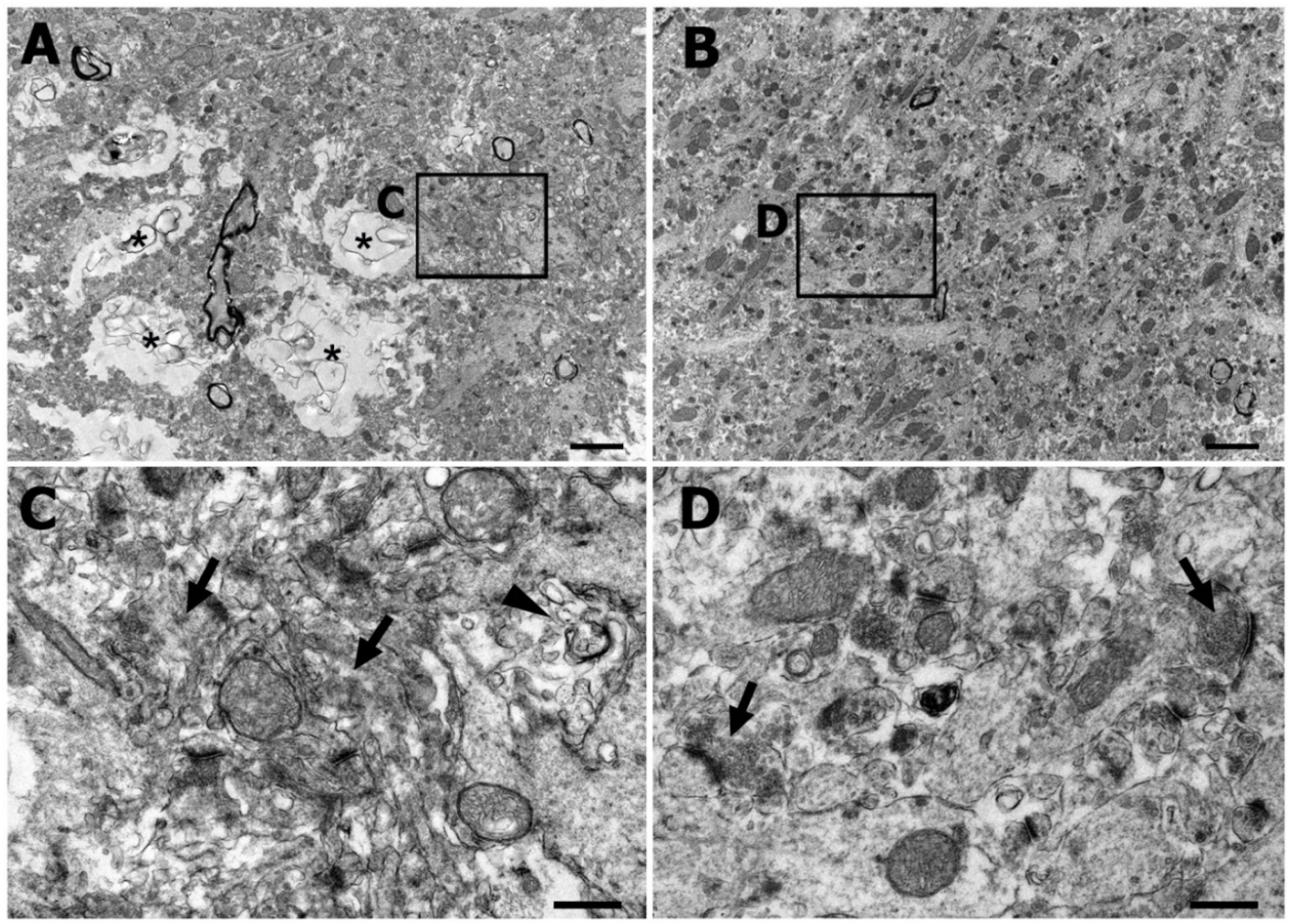
Fallopian tube Semithin section for ciliated epithelium Stained Brightfield  Stock Photo - Alamy
Fallopian tube Semithin section for ciliated epithelium Stained Brightfield Stock Photo - Alamy

IJMS | Free Full-Text | Correlative Light and Electron Microscopy Using  Frozen Section Obtained Using Cryo-Ultramicrotomy
IJMS | Free Full-Text | Correlative Light and Electron Microscopy Using Frozen Section Obtained Using Cryo-Ultramicrotomy

CHAPTER 4 TECHNIQUES Semithin Section Staining with Toluidine Blue 0  CHAPTER 4 TECHNIQUES Semithin Section Staining with Toluidi
CHAPTER 4 TECHNIQUES Semithin Section Staining with Toluidine Blue 0 CHAPTER 4 TECHNIQUES Semithin Section Staining with Toluidi

Applications of Scanning Electron Microscopy Using Secondary and  Backscattered Electron Signals in Neural Structure. - Abstract - Europe PMC
Applications of Scanning Electron Microscopy Using Secondary and Backscattered Electron Signals in Neural Structure. - Abstract - Europe PMC

Electron Microscopy of Synaptonemal Complexes in Semithin Sections Ondina  D. Colman and Juan C. Stockert The ultrastructure of m
Electron Microscopy of Synaptonemal Complexes in Semithin Sections Ondina D. Colman and Juan C. Stockert The ultrastructure of m