Home

slipper Production center Aggregate celery cross section microscope data Constraints Admirable

Vascular bundle of cross section of a celery petiole - UWDC - UW-Madison  Libraries
Vascular bundle of cross section of a celery petiole - UWDC - UW-Madison Libraries

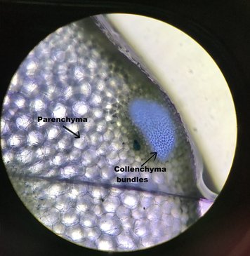
Celery | Cross section showing the Parenchyma and Collenchym… | Flickr

See the Plant Kingdom's Hidden Microscopic Wonders

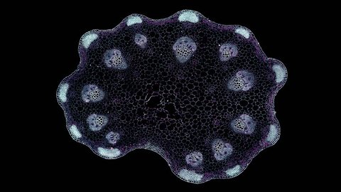
Celery Stripe Cross Section Cut Under Stock Footage Video (100%  Royalty-free) 1016951761 | Shutterstock
Celery Stripe Cross Section Cut Under Stock Footage Video (100% Royalty-free) 1016951761 | Shutterstock
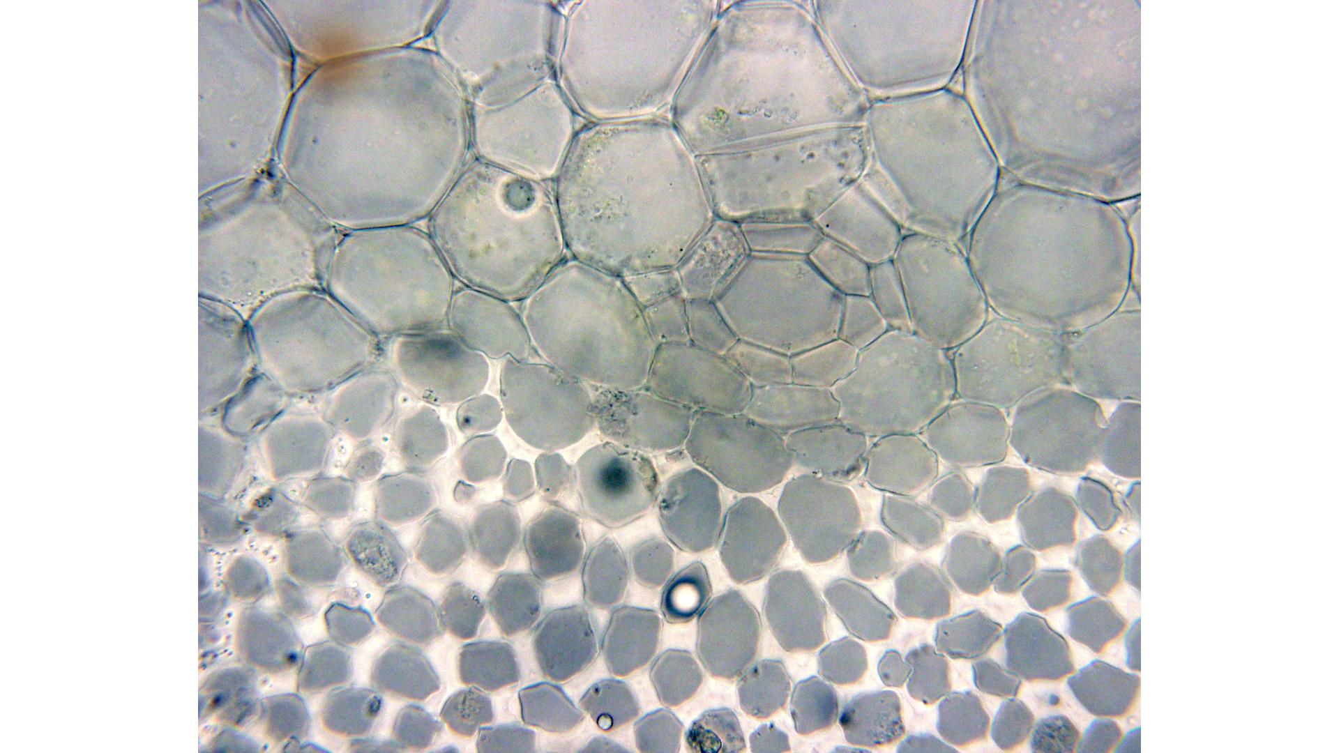
Cross section of a celery petiole - boundary between parenchyma and  collenchyma tissue - 100x objective - UWDC - UW-Madison Libraries
Cross section of a celery petiole - boundary between parenchyma and collenchyma tissue - 100x objective - UWDC - UW-Madison Libraries

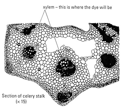
CELERY LAB - Structure and Function of a Plant

Celery Stripe Cross Section Cut Under Stock Footage Video (100%  Royalty-free) 1017144613 | Shutterstock
Celery Stripe Cross Section Cut Under Stock Footage Video (100% Royalty-free) 1017144613 | Shutterstock

Isolation and handedness of helical coiled cellulosic thickenings from  plant petiole tracheary elements | SpringerLink
Isolation and handedness of helical coiled cellulosic thickenings from plant petiole tracheary elements | SpringerLink

File:Celery Cells (400x).jpg - Wikimedia Commons

Observing water moving through plants

Fun Facts About Plant Anatomy | CCBER

General morphology of celery collenchyma (Apium graveolens, eudicot,... |  Download Scientific Diagram
General morphology of celery collenchyma (Apium graveolens, eudicot,... | Download Scientific Diagram
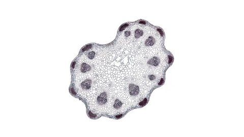
Distribution of chlorophyll in petioles of celery and tobacco.a,... |  Download Scientific Diagram
Distribution of chlorophyll in petioles of celery and tobacco.a,... | Download Scientific Diagram

Category: Celery - OSU Plant Structure (BOT313), Winter 2017

Lab 4: PLANT CELLS, VEGETATIVE ORGAN STRUCTURES, AND PATTERNS OF GROWTH  Flashcards | Quizlet
Lab 4: PLANT CELLS, VEGETATIVE ORGAN STRUCTURES, AND PATTERNS OF GROWTH Flashcards | Quizlet

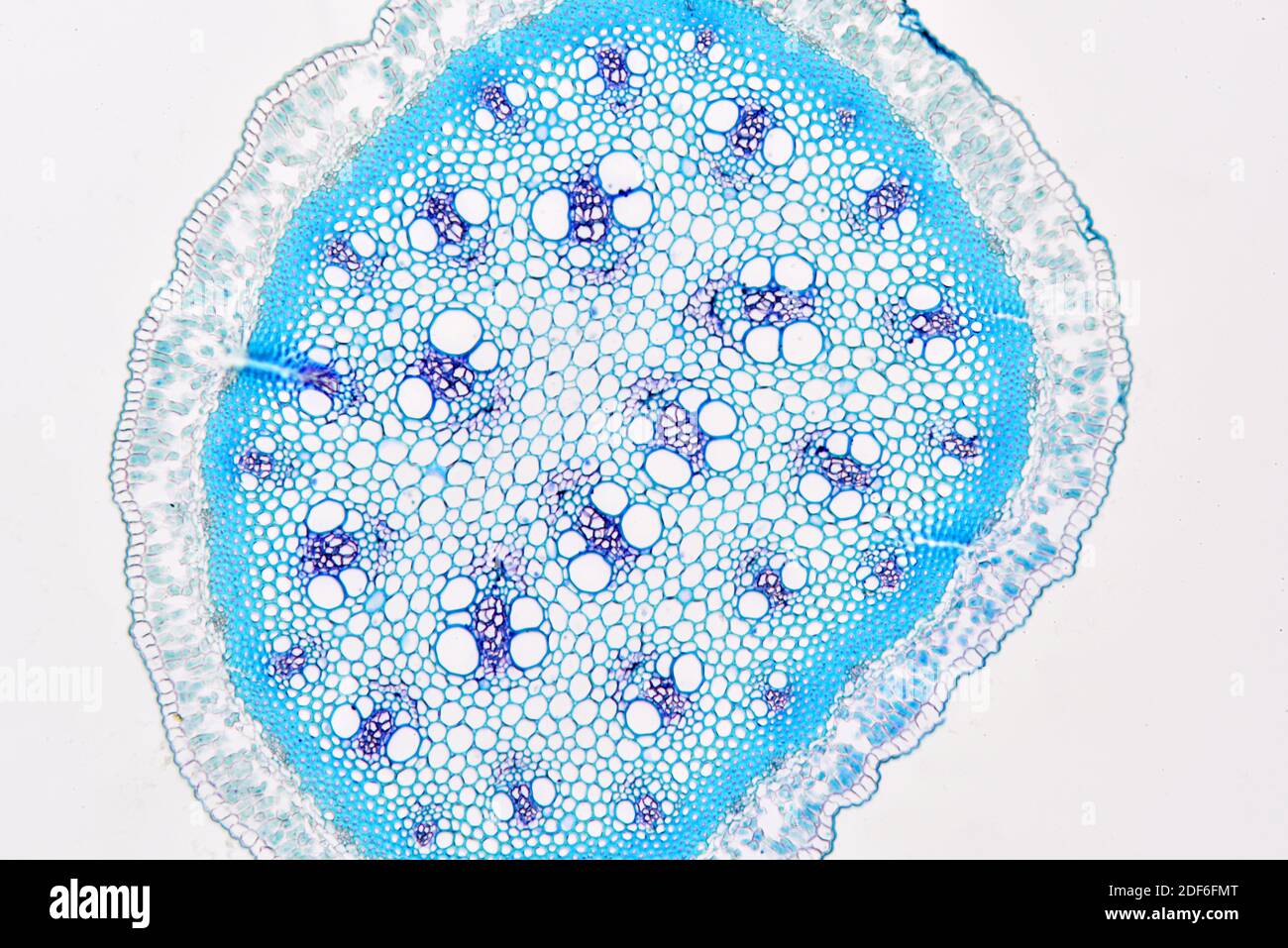
Stem micrograph cross section hi-res stock photography and images - Alamy

Solved 5a.) Now examine the wet mount slides of thin cross | Chegg.com

Plant cells and tissues

The Sorting Hat: An AI-Powered Image Classifier for Cell Biologists | Lab  Manager
The Sorting Hat: An AI-Powered Image Classifier for Cell Biologists | Lab Manager

Celery Stripe Cross Section Cut Under Stock Footage Video (100%  Royalty-free) 1017146368 | Shutterstock
Celery Stripe Cross Section Cut Under Stock Footage Video (100% Royalty-free) 1017146368 | Shutterstock

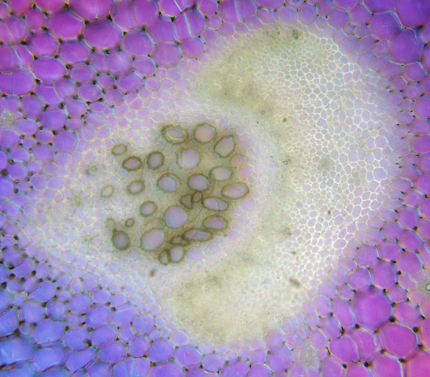
General morphology of celery collenchyma (Apium graveolens, eudicot,... |  Download Scientific Diagram
General morphology of celery collenchyma (Apium graveolens, eudicot,... | Download Scientific Diagram

CELERY LAB - Structure and Function of a Plant

DSC_0237 | Sample: Celery cross section Magnification: 40x S… | Flickr

Celery Lab (Phloem & Xylem) | Biocircuits Outreach

The Microscopic Beauty of Plants and Trees by Robert Berdan - The Canadian  Nature Photographer
The Microscopic Beauty of Plants and Trees by Robert Berdan - The Canadian Nature Photographer

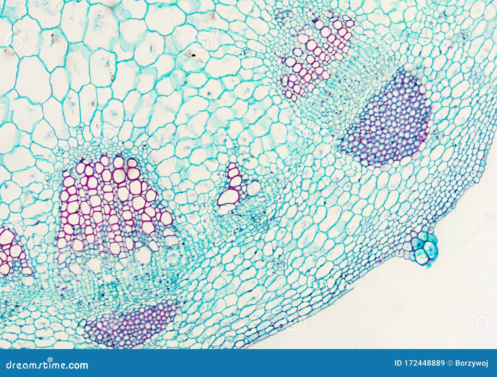
Dicotyledon Stem - Microscopic View Stock Image - Image of micro,  histology: 172448889
Dicotyledon Stem - Microscopic View Stock Image - Image of micro, histology: 172448889